Retinal Detachment
Retinal Detachment in Iran
Best retinal detachment clinic in Iran
Best retinal detachment hospital in Iran
If you’re searching for the best retinal detachment hospital in Iran, choose from our recommended list of top-rated hospitals. These hospitals offer specialized care for retinal detachment, with experienced doctors, advanced technology, and a range of treatment options to ensure the best possible outcome for your procedure. You can expect personalized attention and the highest level of care at these hospitals, which are known for their commitment to patient safety and satisfaction.
- Noor clinic
- Negah clinic
Retinal detachment cost in Iran
There is a significant difference between the cost of the surgery in Iran and other countries. The most important factors for its low price of it in Iran are:
- A large number of clinics for this kind of surgery in Iran
- A large number of applicants for retinal detachment in Iran
Retinal detachment cost in Iran varies depending on the cataract surgery and the hospital. The average cost of the surgery in Iran is $2.800.
Retinal detachment cost in Iran in comparison with other countries
This surgery costs $10.000 in the U.S., $7.300 in Europe, $6.000 in Thailand, and $5.400 in Turkey.
Best retinal detachment surgeon in Iran
One of the most important factors in choosing a good surgeon for this surgery in Iran is the doctor has done many of this kind of surgeries. You can find the best doctors for operation in Iran on our website by following their experiences.
Why should you travel to Iran for a retinal detachment?
Many patients travel to Iran for retinal detachment. One of the reasons for this matter is Iranian specialists and ophthalmologists who have high surgery success rates.
- Low cost of retinal detachment in Iran
- Low cost of accommodation in Iran
- Well experienced doctors
- The high number of retinal detachments in Iran
Diagnostic and surgical centers accordance with today’s European standards is performing the highest quality operations in Iran. Another reason for retinal detachment in Iran is its lower cost compared to other countries.
How long should I stay for retinal detachment in Iran?

About Retinal Detachment
Retinal detachment in Iran or tear repair is eye surgery to place a retina back into its normal position. The retina is the light-sensitive film at the back of the eye. The retina detaches when the retina peels away from the inner wall of the eye. Patients may be more likely to have retinal detachment if they are short-sighted, have had cataract surgery in the past, or have suffered a severe direct blow to the eye. Some types of retinal detachment can run in families, but these are rare. Most retinal detachments occur as a natural aging process in the eye. A detached retina is a serious and sight-threatening event, that happens when the retina becomes separated from its underlying supportive tissue.
The retina cannot function when these layers are detached. And unless the retina is reattached soon, permanent vision loss may result.
- Types of retinal detachment
- Vitrectomy: The retinal tear may have been caused by the vitreous gel (jelly inside the eye) pulling on the blood vessels. In a vitrectomy operation, this gel will be removed from the eye.
- Scleral buckle: the ophthalmologist can help seal any holes in the retina by stitching a piece of silicone rubber or sponge to the outside of the eye. The buckle will not be visible on the outside of the eye. And it will be left in that place permanently.
- Pneumatic retinopexy: In this procedure, the surgeon injects a small bubble of gas into the vitreous body to push the detached portion of the retina onto the retinal pigment epithelium. When a retinal tear or hole hasn’t yet progressed to detachment, the eye surgeon may suggest one of the following procedures to prevent retinal detachment and preserve vision.
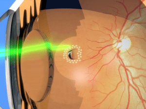
- Laser surgery (photocoagulation). The surgeon directs a laser beam into the eye through the pupil. The laser makes burns around the retinal tear, creating scarring that usually “welds” the retina to underlying tissue.
- Freezing (cryopexy). After giving a local anesthetic to numb the eye, the surgeon applies a freezing probe to the outer surface of the eye directly over the tear. The freezing causes a scar that helps secure the retina to the eyewall. Recommended For:
- Patients that have had cataract surgery in the past or have suffered a severe direct blow to the eye
Before Retinal Detachment
The surgeon will take a medical history from the patient. He or she will run some tests and evaluate the patient’s eyes for more information.
During Retinal Detachment
Patients would be under general or local anesthesia depending on the types of surgery during the procedure. This procedure usually lasts 1 to 2 hours. Patients usually need to stay at the hospital overnight.
Recovery
Patients should place their heads in a specific position so that the gas or oil bubble will lie against the part of the retina that needs support. Patients will need to hold their head in the posturing position for 45 minutes every hour during the day, for five days after the operation; it can be a challenge, but it is just as important as the Retinal detachment in Iran itself. The eyesight may be different after the procedure. The surgeon will explain the kind of results patients can expect, but it may be a few weeks or months before they can tell whether their vision has improved.
Resources: